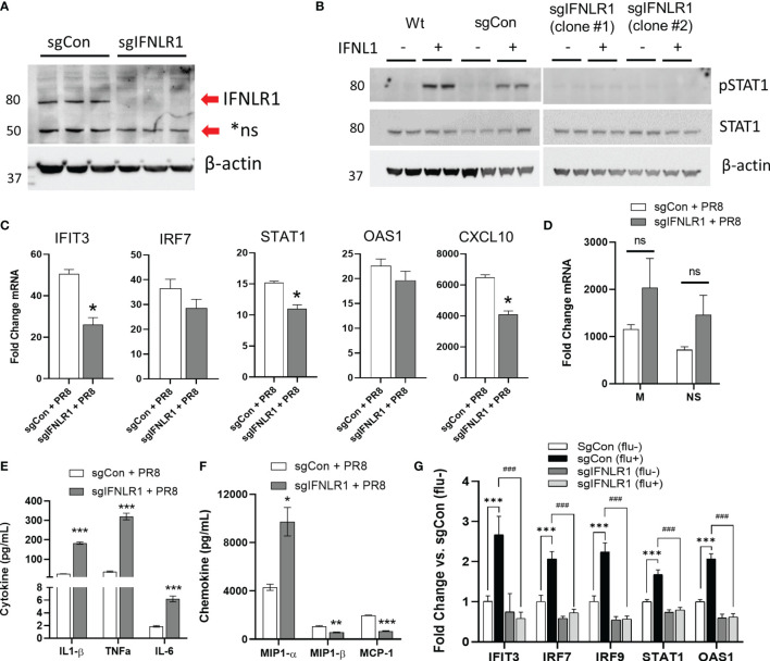
Figure 6.
Macrophage IFNLR1 is indispensible for anti-viral immunity. Control Cas9 THP-1 monocytes (sgCon) and IFNLR1-KO THP-1 monocytes (sgIFNLR1) were differentiated with PMA (20 ng/mL) for 7 days prior to harvest. IFNLR1 protein was measured via immunoblotting (A). sgCon and sgIFNLR1 cells lines were differentiated as described above. At 30 minutes prior to harvest, cells were treated with 50 ng/mL IFNL1. Cells were harvested and total STAT1 and phosphorylated STAT1 induction (pSTAT1) was measured via immunoblotting (B). Representative of 2 independent experiments (A, B). sgCon and sgIFNLR1 were differentiated as above followed by PR8 infection (MOI=0.01) for 24 h. We then measured fold change in ISGs by qRT-PCR. *p < 0.05 vs. sgCon + PR8 by Students T-test (C), fold change in viral mRNA (M gene and NS gene) vs. uninfected (D), and secreted inflammatory cytokines/chemokines in influenza infected macrophages using a custom multiplex from MSD. ns, not significant. *p < 0.05, **p < 0.01, ***p < 0.001 vs. sgCon + PR8 by Multiple T-test with Holm-Sidak correction (E, F). representative of 3 independent experiment, n=3 (C–F). sgCon and sgIFNLR1 THP-1 were differentiated with PMA and treated with supernatants (culture medium) collected from human airway epithelia previously infected with (flu+) or without influenza (flu-) (CA09, MOI=1) ***p < 0.001 vs. sgCon (flu-); ###p < 0.001 vs. sgCon (flu+) by Multiple T-test with Holm-Sidak correction (G).

