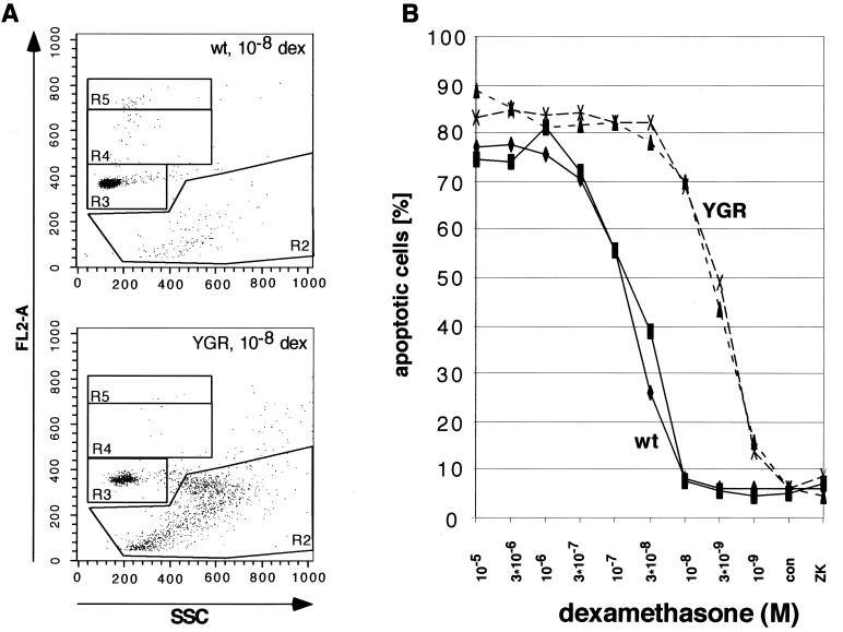
FIG. 5.
Glucocorticoid-induced apoptosis of primary thymocytes of WT (wt) and YGR mice. (A) Flow cytometric analysis of thymocytes cultivated for 9 h in the presence of 10−8 M dexamethasone (dex) and stained with PI. Analysis by gating the cells in region 2 on the basis of their DNA content (FL2-A) and the granulation (SSC) pattern is exemplified. (B) Dose-response curves of dexamethasone-treated thymocytes from four individual mice, two WT and two YGR, are depicted. Cells were cultivated for 9 h in the absence (con) or presence of various concentrations of the GR agonist dexamethasone or after treatment with the GR antagonist ZK112,339 (10−6 M). The degree of apoptosis was determined as described in Materials and Methods and plotted against the concentration of dexamethasone.

