Figure 1.
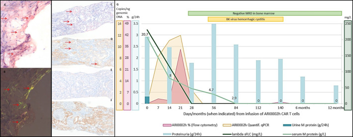
Diagnosis and immunohistochemical typing of amyloidosis. Evolution of clinical and laboratory findings after infusion of ARI0002h. Diagnosis of amyloid deposits in a fine-needle aspiration of subcutaneous fatty tissue: (A) Congo red-stained tissue section, amyloid deposits highlighted with horizontal arrows; (B) Congo red green birefringence, amyloid deposits highlighted with horizontal arrows. Immunohistochemical typing of the renal amyloidosis as ALλ type shown on adjacent sections and amyloid deposits highlighted with horizontal arrows: (C) Congo red-stained (D) anti-ALλ showing positivity on amyloid deposits; (E) anti-ALκ and (F) anti-AA showing negativity on amyloid deposits. Magnification ×100. Amyloid was not evident in the bone marrow section. (G) In the first 6 months after infusion of ARI0002h, a progressive decline in serum M-protein and lambda sFLC was observed. MRD was negative on day +28 and remains negative at 12 months. A renal response with a 55% and 70% decrease in proteinuria was observed at 6 and 12 months, with an intermediate period in which, due to hemorrhagic cystitis, proteinuria is not assessable. Expansion of CARTs by either flow cytometry and qRT-PCR are also represented, with a peak at day +21. AL, amyloid light-chain; CART, chimeric antigen receptor T cell; MRD, minimal residual disease; sFLC, serum free light chain.

