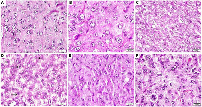
FIGURE 6.
Photomicrograph of an antimesometrial region of the rat uteri at the 8th day of pregnancy (H&E ×400): (A) Control and (B) Control-D400 revealing different sized rounded decidual cells illustrating eosinophilic cytoplasm, vesicular nuclei, and numerous prominent nucleoli. (C) DEF displaying an impaired differentiation of stromal cells into decidual cells. (D) DEF-D400 illustrating alternating decidual cells and impaired differentiated stromal cells (arrows) into decidual cells. (E) DEF-D4000 and (F) DEF-D10000 revealing an obvious differentiation of stromal cells into different sized rounded decidual cells. Scale bar: 20 μm.

