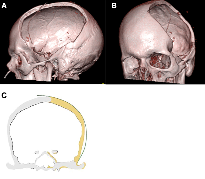
FIG. 1.
(A,B) 3D reconstruction of a CT scan taken post-operatively after decompressive hemicraniectomy. 3D reconstructions were created for all patients and used to measure the surface area of the autologous bone graft. (C) Illustration of a coronal image demonstrating the left cranial defect. 3D, three dimensional; CT, computed tomography.

