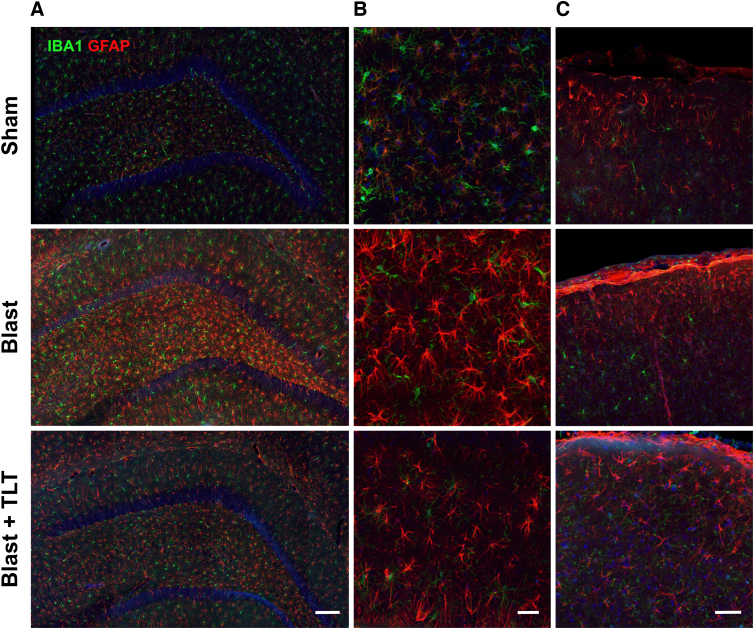
FIG. 7.
Absence of TLT effect on astrogliosis in blast-exposed rats. Sections through the hippocampus (A), hippocampal hilar region (B), or layer I of neocortex (C) immunostained for Iba1 (green) or GFAP (red). In rats exposed to sham TLT, there was increased GFAP staining in the blast-exposed compared to the sham. TLT treatment did not appear to appreciably affect this increased staining. There were no obvious differences in microglial staining (Iba1). Scale bar: 100 (A), 10 (B), and 20 μm (C). GFAP, glial fibrillary acidic protein; Iba1, ionized calcium-binding adaptor molecule 1; TLT, transcranial laser therapy.

