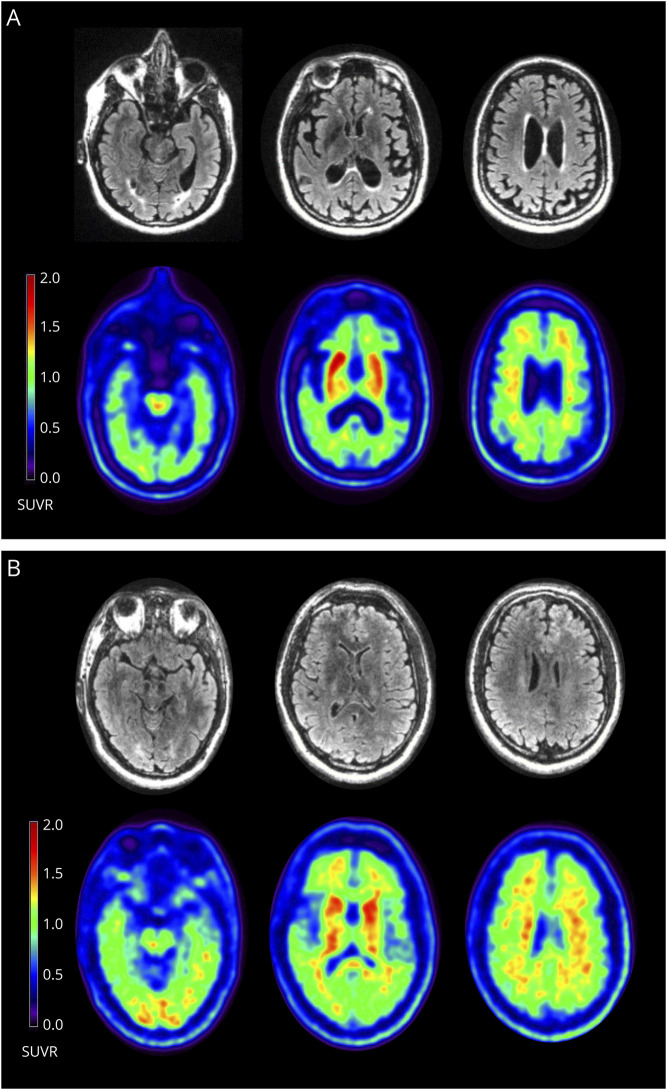
Figure 2. Simultaneous MRI and Amyloid PET in (A) the Proband at Age 45 Years and (B) the Male Cousin at Age 36 Years.
(A) The proband's FLAIR images (first row) demonstrate only trace periventricular white matter changes. The florbetaben PET images (second row) are positive for cortical amyloid and show prominent amyloid deposition in the striatum. The brainstem was used as the reference region for the standardized uptake value ratios (SUVRs). (B) The unaffected male cousin's FLAIR images (third row) are normal. His florbetaben PET images (fourth row) are positive and display similar striatal deposition to (A) that is not seen in older unrelated noncarriers (see eAppendix 4, links.lww.com/NXG/A504).

