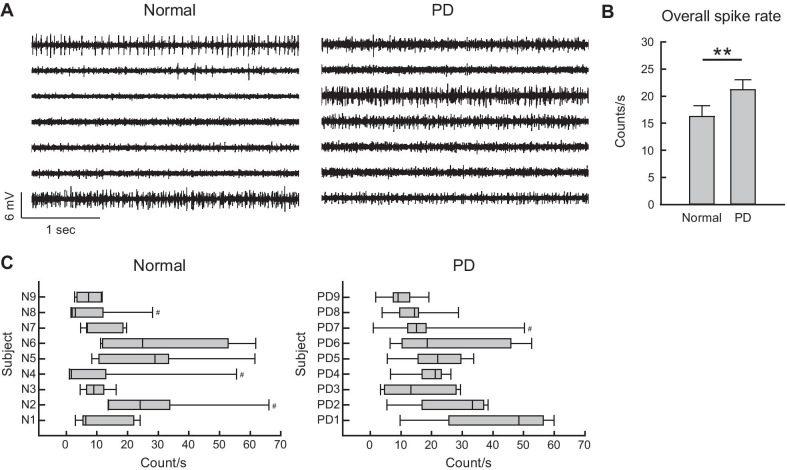
Fig. 2.
Higher MC activities in PD than in normal subjects. A Sample sweeps of MC spikes in a normal and a 6-OHDA hemiparkinsonian (PD) subjects (the left and right panels, respectively). Multi-unit (MU) recordings were obtained from arrays of seven separated insulated tungsten microwires placed in layer V-VI of MC in 8-week-old male Wistar rats. MC discharges at the seven recording sites (from top to bottom) are apparently more diffuse in PD, in comparison to the unevenly distributed pattern in normal subjects (see part C). B A 30-s period of recordings devoid of significant noise from the seven sites were selected for MU analysis for each subject. The sweeps were processed offline by a spike detection software (Sciworks 10.0, Datawave Technology), and the detection threshold was set at 4 times of the estimated median absolute deviation of signals. All detects were included as spikes in multi-unit profiles without further analytic treatment such as clustering. There are higher MU spike rates in PD than in normal subjects (n = 63, or 7 leads in 9 subjects for both groups, **P < 0.01 by Mann–Whitney U test). C Box plots displaying the lower and upper quartiles and the median show the MC spike rate distribution among the seven recording sites in each subject. The whiskers mark the minimum and maximum values of the dataset to display the range of outliers. There are more subjects with box plots skewed rightward in normal than PD subjects, suggesting that the spike rats are less evenly distributed among different leads in normal. #subjects with outliers having individual spike rates > 1.96 standard deviation

