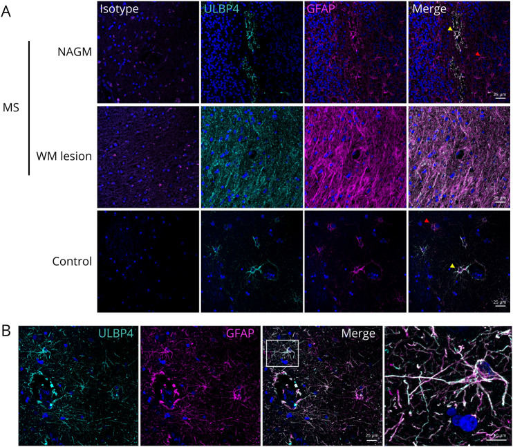
Figure 2. ULBP4 Is Mainly Expressed by Astrocytes in Brain Tissue From Patients With MS and Controls.
(A) Representative images for the costaining of ULBP4 (cyan) and GFAP (magenta) in paraffin-embedded sections from untreated MS donors (NAGM and WM lesion) and controls (no brain related disease); n = 4 for each group. Nuclei were stained with 4',6-diamidino-2-phenylindole (blue). Corresponding isotype controls are shown. Yellow arrows indicate ULBP4+GFAP+ cells, and red arrows indicate ULBP4-negative GFAP+ cells. Scale bars = 25 μM. (B) Representative enlarged image showing colocalization of GFAP and ULBP4 in the brain section. Scale bars = 10 µM. GFAP = glial fibrillary acidic protein; MS = multiple sclerosis; NAGM = normal-appearing gray matter; WM = white mater; ULBP4 = UL16-binding protein 4.

