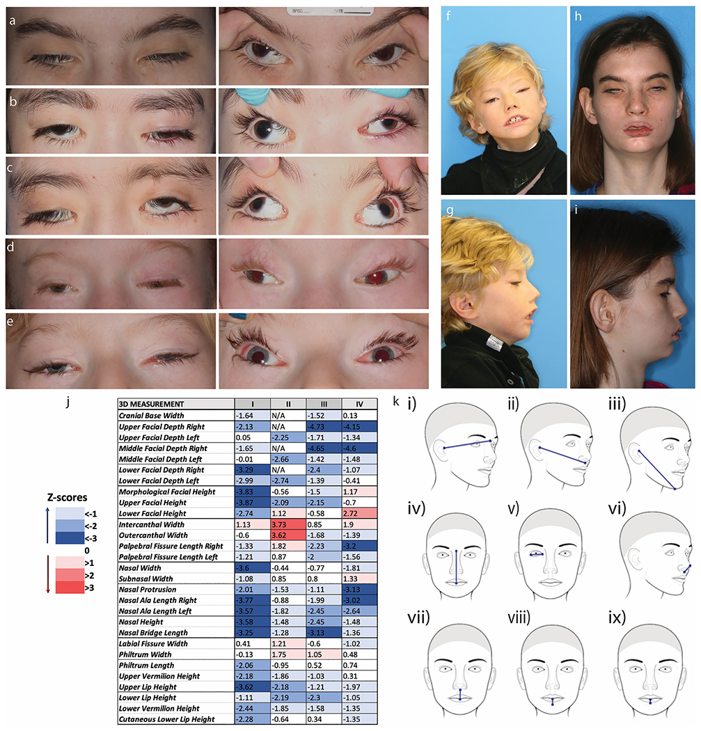
Fig. 1. Ophthalmological and Craniofacial Evaluation.

All subjects have severe ptosis, extremely limited eye movements, and severe exotropia. a-e Ocular photographs of the five subjects evaluated at NIH (XIII, X, XI, XII, VII). Panels on the left show the severe ptosis. Panels on the right, with the lids artificially raised, display the severe exotropia. Subjects have almost no voluntary eye movements. F-I. Frontal and profile photos of subjects VII (f&g) and XIII (h&i). j. Color scale table depicting the deviation of the soft-tissue measurements in four TUBB3 R262H subjects, expressed in the form of Z-scores, which were computed from the normal values provided by FaceBase. k. Schematic representation of the most negatively affected linear 3D measurements in this cohort. In general, most values are lower than the average normal values, particularly the facial depth measures (i, ii, iii), the upper facial height (iv), the palpebral fissure lengths (v), the protrusion of the nose (vi), and the vertical dimensions of the lips (vii, viii, ix). The only measure with increased or normal values in all four subjects is the intercanthal width.
