Figure 3.
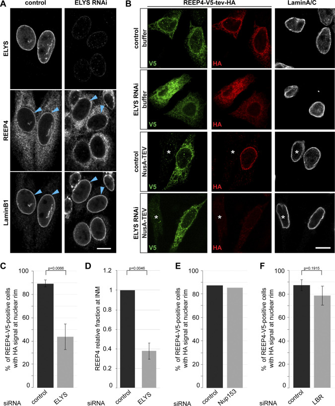
ELYS promotes REEP4 INM targeting. (A) REEP4-HA cells treated with control or ELYS-targeting siRNA; labeled for the HA tag, ELYS, and Lamin B1; and imaged by confocal microscopy. Arrowheads indicate the nuclear rim. (B–D) NusA-TEV assay of HeLa cells expressing REEP4-V5-tev-HA, treated with control or ELYS siRNA, and stained for V5 tag, HA tag, and Lamin A/C. (B) Representative images. Asterisks indicate untransfected cells. (C) Quantification of cells with HA staining at the nuclear rim after NusA-TEV treatment (n = 5). (D) For an estimate of changes to the fraction of REEP4 at the nuclear rim, HA signal intensity (i.e., INM pool) was divided by V5 signal intensity (i.e., total pool of REEP4), and the mean value was normalized to the HA/V5 ratio in the respective control. n = 5 with 25 cells analyzed per condition. (E and F) Fraction of REEP4-V5-tev-HA–expressing, NusA-TEV–treated cells with HA staining at the nuclear rim in control cells or after depletion of Nup153 (E; average of two experiments with very similar outcomes) or LBR (F; n = 3). In A and B, scale bar is 10 µm. In C, E, and F, ≥35 cells were analyzed per condition in a blinded manner. In C, D, and F, error bars are SEM. P values were obtained using two-tailed, paired t tests. See Fig. S1, A–D, for analysis of ELYS, Nup153, and LBR depletions and expression of REEP4-V5-tev-HA.

