Figure 4.
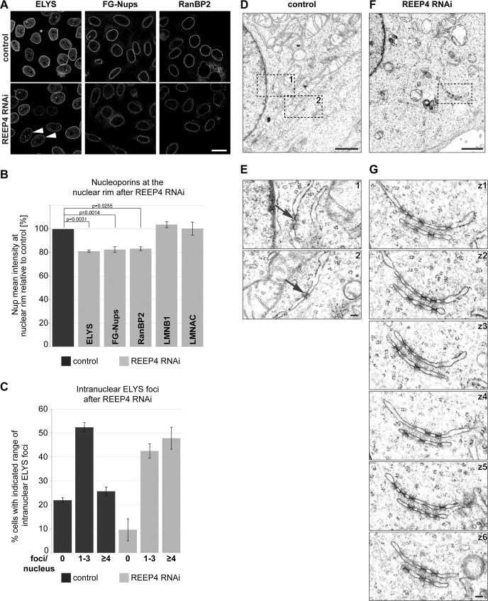
REEP4 is required for normal NPC densities. (A–C) HeLa cells treated with control or REEP4 siRNA, immunolabeled for indicated nucleoporins, and imaged by confocal microscopy. (A) Representative images. Arrowheads indicate cells with increased nucleoplasmic ELYS foci. (B) Mean intensities of the respective nuclear rim proteins in control and depleted cells. Lamin B1 (LMNB1), Lamin A/C (LMNAC), n = 3; ELYS, RanBP2, n = 4; FG-Nups, n = 6. At least 100 cells were analyzed per condition. Error bars are SEM. Two-sided, paired t tests were performed on the raw data and yielded the indicated P values. (C) Percentage of cells with zero, one to three, or four or more intranuclear ELYS foci in control and REEP4 RNAi cells. n = 3; ≥55 cells were analyzed per condition in a blinded manner; error bars are SEM. (D–G) Transmission EM analysis of control and REEP4 RNAi cells. (D and F) Overview images. (E) Enlarged views of the regions marked as 1 and 2 in D. Arrows indicate examples of rare NPC intermediates in the ER of control cells. (G) Images of consecutive serial z-sections (z1–z6) of the region outlined in F, showing clustered NPC intermediates in stacked ER cisternae in the cell periphery in REEP4 RNAi cells. See Fig. S2 G for another example. Scale bars: 20 µm (A), 1 µm (D and F), 100 nm (E and G).

