Figure S1.
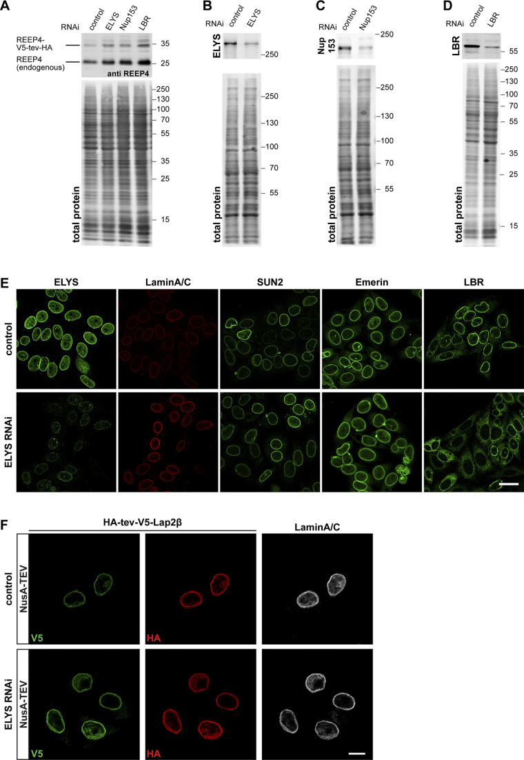
Role of ELYS in REEP4 INM targeting. Related to Fig. 3. (A) Cells transfected with a construct for expression of REEP4-V5-tev-HA and with control, ELYS-, Nup153-, or LBR-targeting siRNA were lysed in Laemmli buffer and analyzed by SDS-PAGE and immunoblotting using an antibody against REEP4, which detects both endogenous REEP4 and REEP4-V5-tev-HA. Expression of REEP4-V5-tev-HA was not reduced after either of the siRNA treatments. (B–D) Lysates of cells transfected for the TEV assays with nontargeting control and ELYS- (B), Nup153- (C), or LBR- (D) targeting siRNA were analyzed by SDS-PAGE and immunoblotting with antibodies against the respective depleted proteins. (B and C) ELYS (n = 4) and Nup153 (n = 2) protein levels were on average reduced by 70%. (D) LBR protein levels were on average reduced by 90% (n = 3). (A–D) Images below the immunoblots show total protein stain of the membrane to visualize the amounts of loaded protein in the different samples. The total protein amounts in the respective lanes were used for normalization before determining depletion efficiency. (E) HeLa cells were treated with control or ELYS siRNA and immunostained for ELYS and the INM proteins Lamin A/C, SUN2, Emerin, and LBR. After ELYS-specific RNAi, ELYS was markedly reduced at the nuclear rim (far left column) but localization of the INM proteins Lamin A/C, SUN2, and Emerin was not impaired. Lamin A/C staining appeared increased after ELYS depletion for unknown reasons. Note that the two left columns show the same cells for control and ELYS RNAi, respectively, that were labeled for both ELYS and Lamin A/C. LBR targeting to the INM was impaired after ELYS depletion (far right column), as previously described by Mimura et al. (2016). Scale bar is 20 µm. (F) Cells expressing the INM protein Lap2β tagged N-terminally with HA-tev-V5 were treated with control or ELYS-targeting siRNAs and subjected to NusA-TEV treatment. Only the NusA-TEV treatment condition is shown; buffer-treated cells show an identical pattern. At least 30 cells were analyzed per condition in two different experiments. All control cells with V5-labeling, indicating successful transfection, also showed HA-staining of corresponding intensity. Among 68 V5-positive ELYS RNAi cells, one cell was lacking clear HA staining, but all other cells showed HA staining that corresponded to V5 intensity, suggesting that ELYS depletion did not lead to increased permeability of NPCs for NusA-TEV protease. Scale bar is 10 µm.

