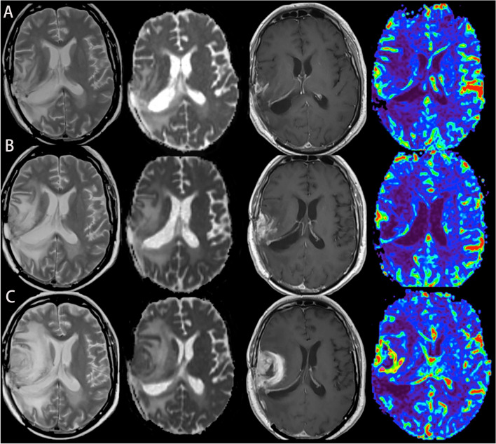
Figure 1.
A case of glioblastoma relapsed during immunotherapy, T2, ADC map, T1-enhanced, and CBV map from left to right. (A–C) MRI was performed in the 2nd, 6th, and 8th months of immunotherapy, respectively, showing that the edema degree of the lesion was gradually aggravated, the enhancement was more obvious, and the perfusion was higher.

