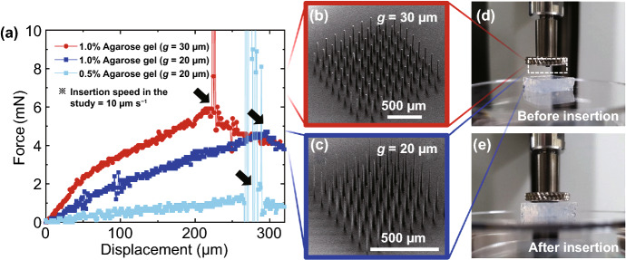
Fig. 9.
Insertion tests of the fabricated microneedle arrays to agarose gels mimicking brain tissues. a Force–displacement response during the insertion of microneedle arrays into 0.5% and 1% agarose gels at an insertion rate of 10 μm s−1. b, c SEM images of the microneedle arrays used in the insertion tests, which were fabricated from 30 and 20 μm gap sizes (for b and c, respectively). d, e Photographs of the experiments taken before (d) and after (e) insertion of a microneedle array

