Figure 4.
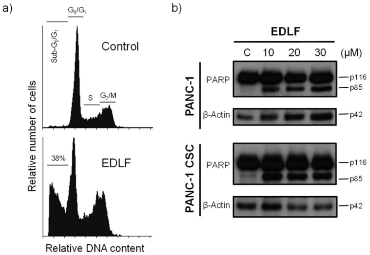
Induction of apoptosis in PANC-1 CSCs by edelfosine. (a) PANC-1 CSCs were incubated in the absence (control) or presence of 20 μM edelfosine (EDLF) for 5 days, and then apoptosis was analyzed by means of flow cytometry. The different cell cycle stages are indicated. The percentage of apoptotic cells in the sub-G0/G1 region is shown. The data are representative of at least three independent experiments. (b) PANC-1 cells and PANC-1 CSCs were untreated (C) or treated with edelfosine (EDLF) at the indicated concentrations for 48 h and analyzed by means of Western blotting using specific antibodies for PARP; β-actin was used as the loading control. Molecular weights (in kilodaltons) of every protein are indicated at the right side of each panel. The gels were cropped to show the relevant sections. The Western blot images are representative of three independent experiments.

