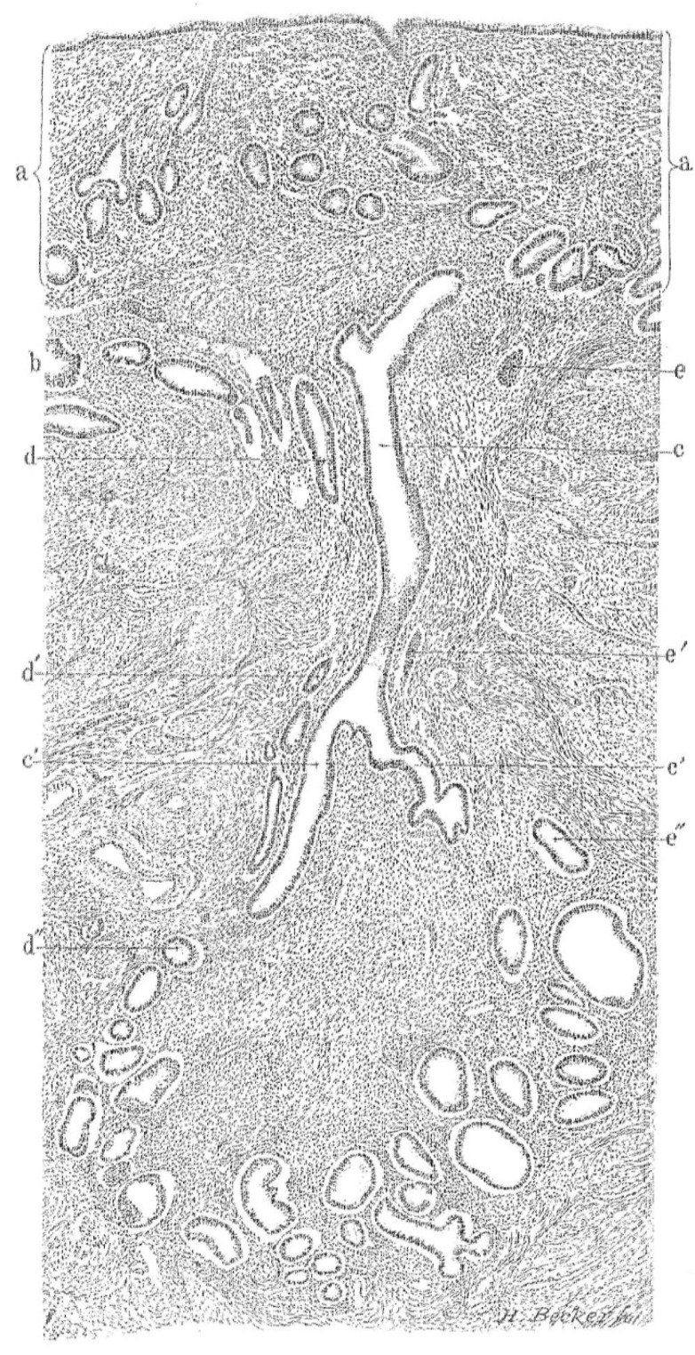
Figure 1.
Histological section of the uterus with adenomyosis. The gland (c) within the muscle (b) is in continuity with the mucosa (a). The section also demonstrates gland continuity and convolutions deep within the muscle layer. In this illustration (c,c’) represent one gland and its branches and (d,d’,d’’) and (e,e’,e’’) represent adjacent glands. The illustration demonstrates the meticulous work by Thomas Cullen (1908).

