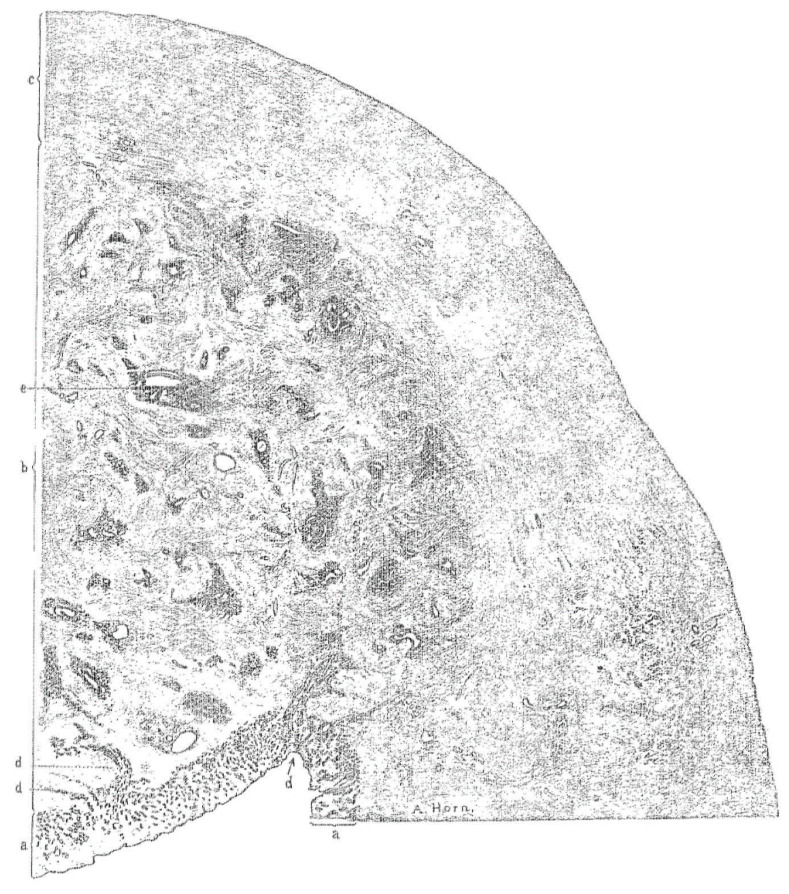
Figure 2.
Section of the upper half of the posterior wall of the uterus with adenomyosis. The wall has three distinct zones: (a) Inner mucosa, (b) region with adenomyosis, (c) outer normal muscle tissue. The gland at (d) extends directly into the muscle and at (e) the gland is retracted from the surrounding stroma. The illustration demonstrates the meticulous work by Thomas Cullen (1908).

