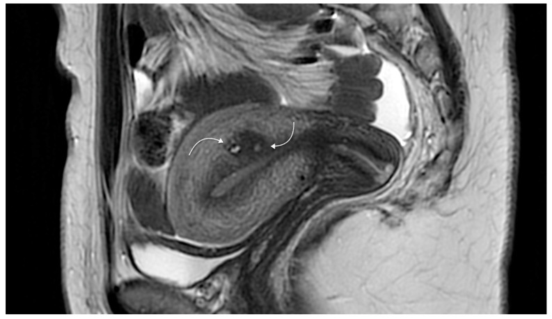
Figure 4.
Magnetic resonance imaging showing sagittal two-dimensional T2-weighted magnetic resonance images showing a hypointense lesion containing tiny spots (curved arrows) and located in the posterior wall, adjacent to the endometrial cavity related to focal adenomyosis. Reproduced with permission from Habiba et al. (2020).

