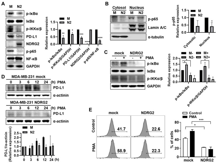
Figure 2.
The inhibitory effects of NDRG2 on PD-L1 expression through NF-κB signaling. (A) Western blotting was performed with MDA-MB-231 mock or NDRG2 whole cell lysates to measure p-IκBα, IκBα, NF-κB, p-IKKα/β, p-p65, PD-L1, and NDRG2 expression. (B) The cell lysates were fractionated into nuclear and cytosolic compartments to assess p-p65 expression levels. (C) Cells were treated with 40 ng/mL of PMA for 1 h to measure the p-IκBα and p-IKKα/β expression levels. (D) PMA was administered for the indicated times to evaluate the effect of PMA on PD-L1 expression, and Western blotting was performed with whole-cell lysates. (E) After 12 h of PMA stimulation, the cells were harvested, and the PD-L1 expression on the surface of MDA-MB-231 cells was measured by flow cytometry. All experiments were independently repeated at least 3 times. * p < 0.05, ** p < 0.01, *** p < 0.001, and **** p < 0.0001. The uncropped blots and molecular weight markers are shown in Supplementary Materials.

