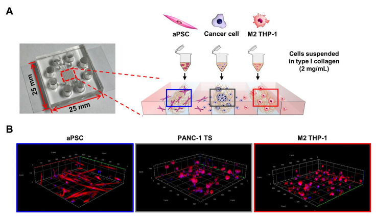
Figure 1.
Schematic of the 3D co-culture system using a microfluidic channel chip. (A) Cells were pre-conditioned and suspended in type I collagen solution (2 mg/mL). Cells were then loaded into each designated cell channel of the microfluidic channel chip. (B) 3D reconstruction images of PANC-1 TSs, aPSCs, and M2 THP-1 cells cultured in collagen matrix. See Materials and Methods for details. TS: tumor spheroid; aPSC: activated pancreatic stellate cell; M2 THP-1: THP-1-derived M2 macrophage.

