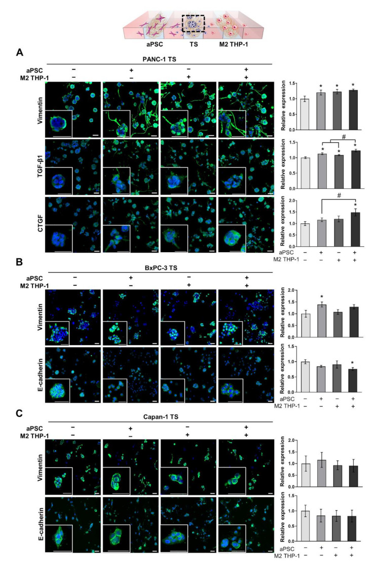
Figure 3.
Increased expression of EMT-related proteins in pancreatic TSs under 3D co-culture conditions with stromal cells. (A) Expression level of vimentin, TGF-β1, and CTGF in PANC-1 TSs with or without aPSCs, M2 THP-1 cells, or both. (B,C) Expression level of vimentin and E-cadherin, which are representative EMT markers, in BxPC-3 and Capan-1 TSs with or without aPSCs, M2 THP-1 cells, or both. Cells were counterstained with DAPI. aPSCs and M2 THP-1 cells were pre-conditioned before loading into the microfluidic channels. Data represent the mean ± SD of three independent experiments. Scale bar: 50 µm, * p < 0.05 compared to TSs cultured alone. # p < 0.05 compared to TSs co-cultured with either aPSCs or M2 THP-1 cells. Cells were grown for 5 days before analysis.

