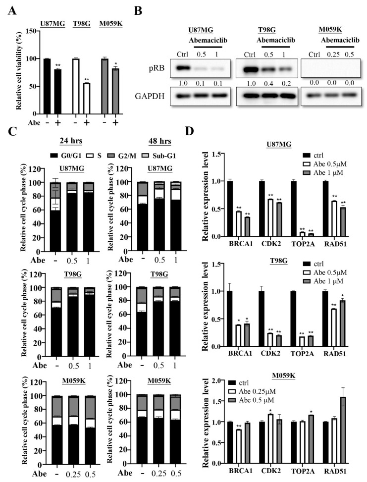
Figure 1.
Abemaciclib treatment regulates cell proliferation on the U87MG, T98G and M059K cells. (A) The three cancer cells were treated with abemaciclib for 72 h and the proliferation rate was evaluated by using MTT assay. Data are presented as the mean ± SD of duplicate wells, and are representative of two independent experiments. Abe: abemaciclib. (* p < 0.05, ** p < 0.01 compared to control group) (B) The three cancer cells were treated with the indicated concentrations (μM) of abemaciclib for 24 h and the levels of phosphorylated RB were detected via Western blot analysis (Figure S1). (C) The three cancer cells were exposed to the indicated concentrations (μM) of abemaciclib for 24 and 48 h and then subjected to cell cycle analysis. Data are presented as the mean ± SD (n = 2). (D) Expressions of indicated genes in the three cancer cells were validated by qPCR. The results are presented as the mean ± SD for duplicate samples. (* p < 0.05, ** p < 0.01 compared to control group).

