Figure 3.
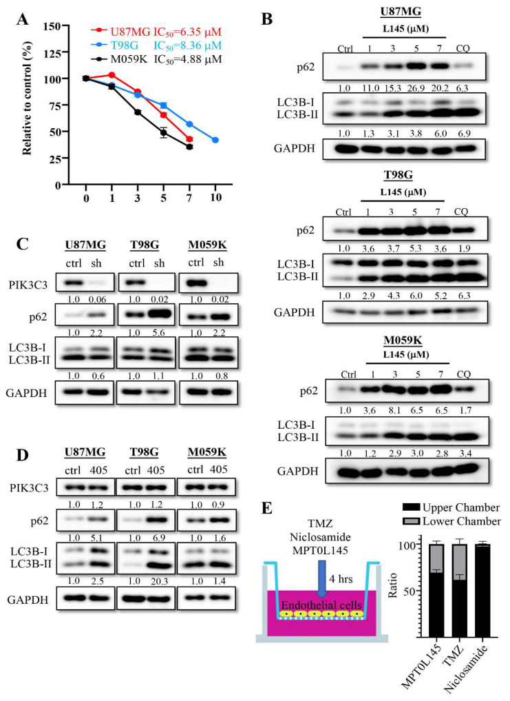
MPT0L145 increases incomplete autophagy in U87MG, T98G and M059K cells. (A) The three cancer cells were treated with MPT0L145 for 72 h and the IC50 value was evaluated using MTT assay. Data are presented as the mean ± SD of triplicate wells, and are representative of two independent experiments. (B) The three cancer cells were exposed to the indicated concentrations (μM) of MPT0L145 and chloroquine (12.5 μM) for 24 h. The protein lysates were subjected to Western blot analysis with the indicated antibodies (Figure S3). (C) PIK3C3 was stably depleted in the three cancer cells and the levels of indicated proteins were detected via Western blot analysis (Figure S3). (D) The three cancer cells were exposed to the PIK3C3 inhibitor SAR405 (10 μM), for 24 h. Protein lysates were subjected to Western blot analysis with the indicated antibodies (Figure S3). (E) TMZ, Niclosamide and MPT0L145 were injected into the upper chamber of the ready-to-use BBB kit for 4 h. The supernatants of the upper and lower chambers were collected and analyzed by LC/MS/MS. The ratio was calculated as the concentration of indicated chamber/(concentration of lower chamber plus concentration of upper chamber) × 100% and the data are presented as the mean ± SD of four trans wells.

