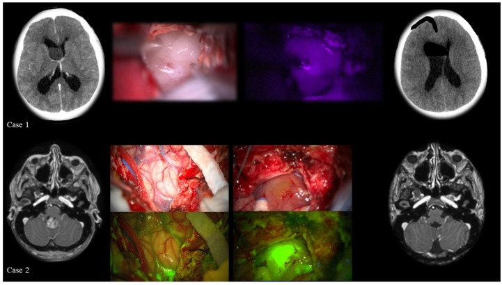
Figure 2.
Supratentorial (case 1) and infratentorial (case 2) ependymomas. Case 1: a 50-year-old female patient with a lesion in the frontal horn of the right lateral ventricle: a right fronto-parietal transcortical approach was used to remove the tumor whose histology, based on the WHO classification, was A grade I ependymoma). From left to right: pre-operative CT scan; intraoperative image of the tumor under white light microscope illumination; blue light illumination using 5-ALA: in this case the tumor was not fluorescent; and finally, the post-operative CT scan. Case 2: a 29-year-old female patient with an intraventricular lesion in the IV ventricle, with moderate contrast enhancement in T1-weighted MRI scan. The histological diagnosis revealed a grade III ependymoma (WHO 2016).

