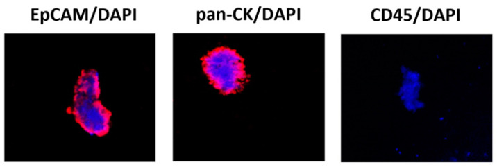
Figure 1.
Confocal microscopy representative images of isolated CTCs stained for epithelial and hematopoietic markers. Representative image by confocal microscopy of circulating cancer cells stained with anti-human EPCAM, anti-human pan-CK or anti-human CD45 Abs followed by goat anti-mouse secondary antibody Alexa 594-conjugated. DAPI was used to counteract nuclei. Magnification 100×. CK = cytokeratin, an epithelial cytoplasmic marker; DAPI = nuclear marker; EpCAM = epithelial membrane marker; CD45 = leukocyte common antigen.

