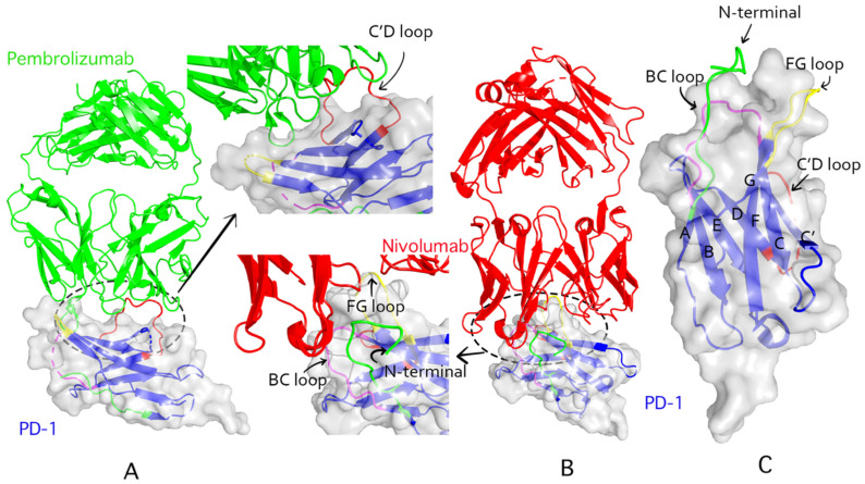
Figure 2.
Structural interactions of pembrolizumab and nivolumab with PD-1. (A): Pembrolizumab complexed (PDB ID: 5jxe) with human PD-1 extracellular domain. The light green ribbons represent pembrolizumab. PD-1 is represented by the transparent blue ribbons. (B): The complex of nivolumab (PDB ID: 5wt9) and PD-1 extracellular domain; the nivolumab is represented by red ribbons. (C): The extracellular domain of human PD-1 (PDB ID: 3rrq). The BC loop, C’D loop, FG loop, and N-terminal domain are in magenta, red, yellow, and green colors, respectively.

