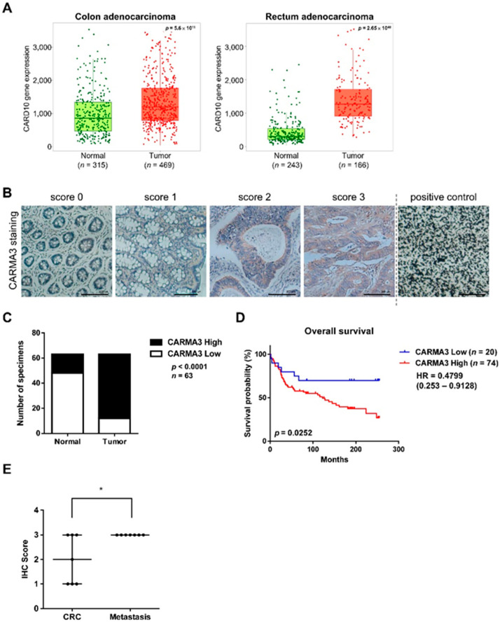
Figure 1.
Higher expression of CARMA3 correlates with malignant progression of colorectal cancer (CRC). (A) Boxplot of CARMA3 gene expression in the colon (left) and rectum adenocarcinoma (right) were determined for comparing paired normal and tumor samples, which were downloaded from TNMplot. The dataset was analyzed by the Mann–Whitney U test. (B) CARMA3 immunohistochemical (IHC) staining in the colon and CRC TMA tissues with scores of 0–3 was shown. Carbon was used as a positive control. Magnification: 200×. Scale bar, 100 μm. (C) Quantification for IHC staining of CARMA3 in paired specimens of CRC and adjacent normal colon tissues was shown. The Chi-square analysis followed by Fisher’s exact test was used to analyze the significant difference. (D) Kaplan–Meier analysis of the overall survival of 94 colorectal cancer patients in TMA with low and high expression of CARMA3 (p = 0.0252, log-rank test, HR = 04799) was shown. CARMA3 expression was classified according to the median of the IHC score of specimens. (E) CARMA3 expression positively correlated with metastatic tissues compared to paired specimens of CRC tissues in TMA (n = 7). *, p < 0.05 by paired two-tailed Student’s t-tests.

