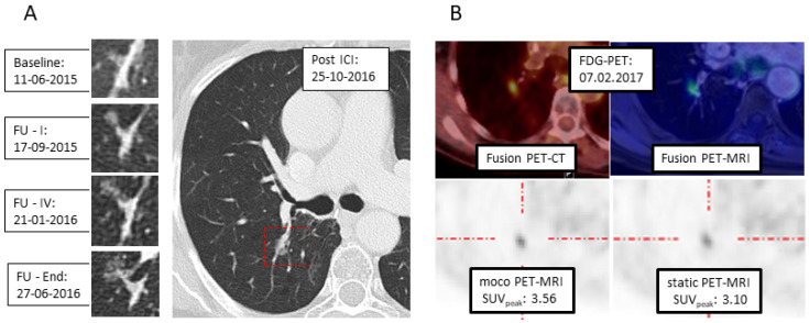
Figure 2.
Example of respiratory motion correction in PET-MRI. (A) CT: Development of a semi-solid formation post-immunotherapy at the right hilum. (B) 18F-FDG-PET with PET/CT (upper left) und PET-MRI (upper right): With MRI-based motion correction (moco), a higher SUVpeak is calculated (lower left), compared to static reconstruction (lower right).

