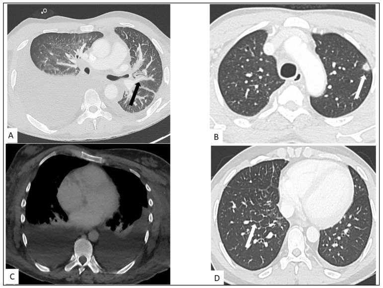
Figure 4.
Thoracic involvement in KS. (A) Highly abundant right pleural effusion and low abundance left pleural effusion with peribronchovascular thickening (arrow). (B) Nodule with halo sign in the left superior lung (arrow). (C) Bilateral pleural effusion on contrast-enhanced CT-scan. (D) Interlobular septal thickening (arrow) in a secondary pulmonary lobule of middle and lower lungs.

