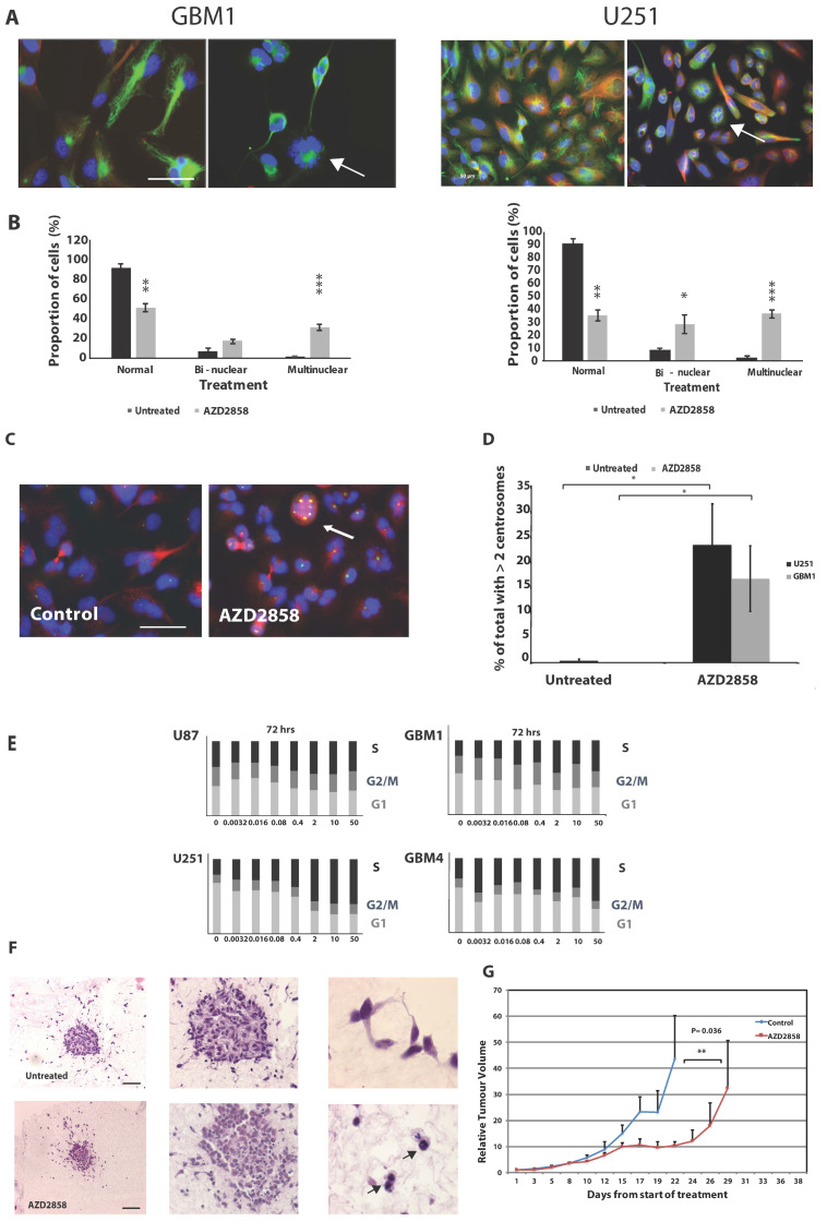
Figure 2.
Treatment with AZD2858 induces a multinuclear phenotype in patient-derived stem cell-like lines and established glioma cell lines. (A) Cell lines were treated with AZD2858 at 1 µM concentration and allowed to incubate for 72 h. They were then fixed with 100% methanol and stained with anti-tubulin antibody (green) and DAPI (blue). Representative images by confocal microscopy are shown for the cell line GBM1 and U251. The arrow indicates multinucleation. Scale bar = 20 µm. (B) Immunofluorescence images were used for counting and quantification of cells for the presence of two (bi) or more (multi) nuclei per cell. At least 200 cells/cell line were scored. Results are shown for the patient-derived stem cell-like cell line GBM1 and the established cell line U251. (C) Cell lines were treated with AZD2858 at 1 µM concentration and allowed to incubate for 72 h. They were then fixed with 100% methanol and stained with anti-pericentrin antibody (green) and DAPI (blue). Representative images by confocal microscopy are shown for the cell line U251. The arrow indicates the presence of several centrosomes. Scale bar = 20 µm. (D) Immunofluorescence images were used for counting and quantification of cells for the presence of two or more centrosomes/cell. At least 200 cells/cell line were scored. (E) Established (U87, U251) and patient-derived stem cell-like cell lines (GBM1, GBM4) were mock-treated or treated with AZD2858 at 5-fold dilutions from 50 to 0.0032 µM and prepared for FACS analysis. S-phase arrest following exposure to AZD2858 was recorded after 72 h exposure in established and patient-derived stem cell-like cell lines at doses > 1 µM. (F) GBM4 3D spheroids embedded in collagen were treated with 500 nM AZD2858 for 72 h and then fixed with 4% paraformaldehyde for immunohistochemistry labeling. Glioma spheroids maintained within the original collagen plug were embedded in paraffin, sectioned, and then stained with H&E to visualize cellular features including the presence of bi- or multinucleated cells within the spheroids. Cells with enlarged nuclei and atypical nuclei number are indicated by the arrow (magnification left to right ×4, ×10, and ×40); scale bar = 50 µm. (G) In a U87 subcutaneous model, 1 × 10⁶ U87-MG cells were injected subcutaneously into the right flank of BALB/c Nude mice. Once tumors were palpable (approximately 5 mm diameter), animals were randomly assigned into experimental groups. AZD2858 was given at 10 mg/kg/day × 10 doses following fractionated irradiation (3 × 5 Gy) and tumor volumes were assessed. A significant effect on growth delay, p < 0.01 for AZD2858 treatment versus control was noted beyond day 17 (**). * denotes p ≤ 0.05, ** denotes p ≤ 0.01 and *** denotes p ≤ 0.001.

