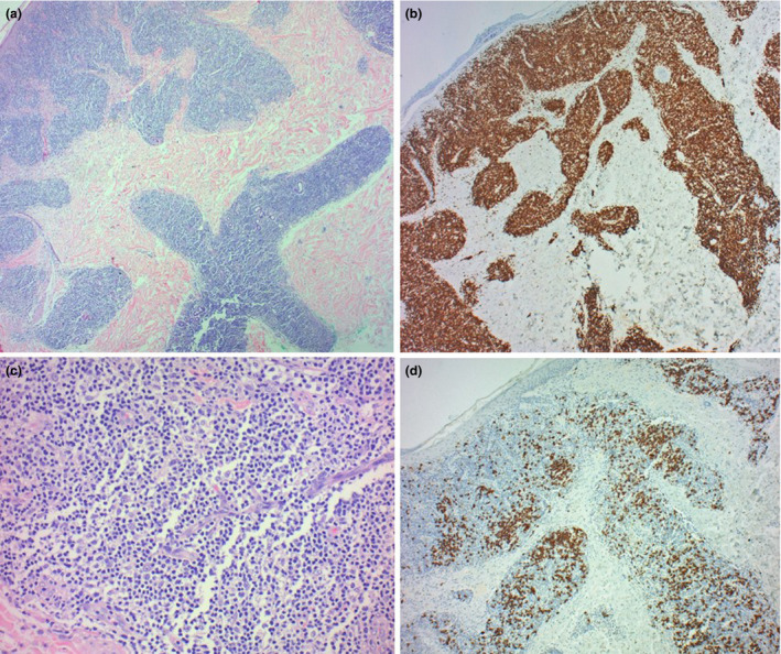
Figure 2.

(a) Nodular lymphoid infiltrate extending from the superficial dermis to the deep dermis, with a focal interface at the basal layers of the epidermis. (H&E, x100). (b) Polymorphous infiltrate with a predominance of small mature lymphocytes. (H&E, x 200). Immunohistochemistry shows a mixture of B and T cells, highlighted by CD3 (c) and CD20 (d) respectively, with a predominance of T cells (x100).
