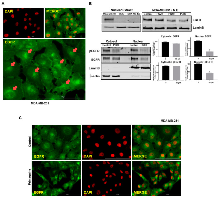
Figure 3.
nEGFR expression was reduced in MDA-MB-231 cells upon primaquine treatment. (A) Nuclear localization of EGFR with anti-EGFR (green) and nuclei (red, DAPI) using a fluorescence microscope (Lionheart FX, BioTek, Winooski, VT, USA). nEGFR resulted from merging of EGFR (green) and DAPI (red). (B) MCF-7, MDA-MB-453, and MDA-MB-231 cells were treated with primaquine (20, 40, and 80 μM) for 24 h and subjected to Western blot analysis. Cells were finally lysed, and cytosolic and nuclear proteins were isolated. The cytosolic and nuclear proteins were identified with anti-pEGFR and anti-EGFR antibodies by Western blotting. The expression of lamin B and β-actin were determined as a loading control for nuclear and cytosolic lysates. Values are the mean ± SD of 3 independent experiments. * indicates p < 0.05 vs. control. (C) Cancer cells with/without primaquine were washed, fixed, permeabilized, and blocked with 0.1% normal goat serum for 60 min. Cells were incubated with primary monoclonal EGFR antibody. Immunostained cells were examined with a fluorescence microscope (Lionheart FX, BioTek, Winooski, VT, USA). Red, nuclei stained by DAPI; green, EGFR. nEGFR was identified by merging EGFR (green) and DAPI (red) images.

