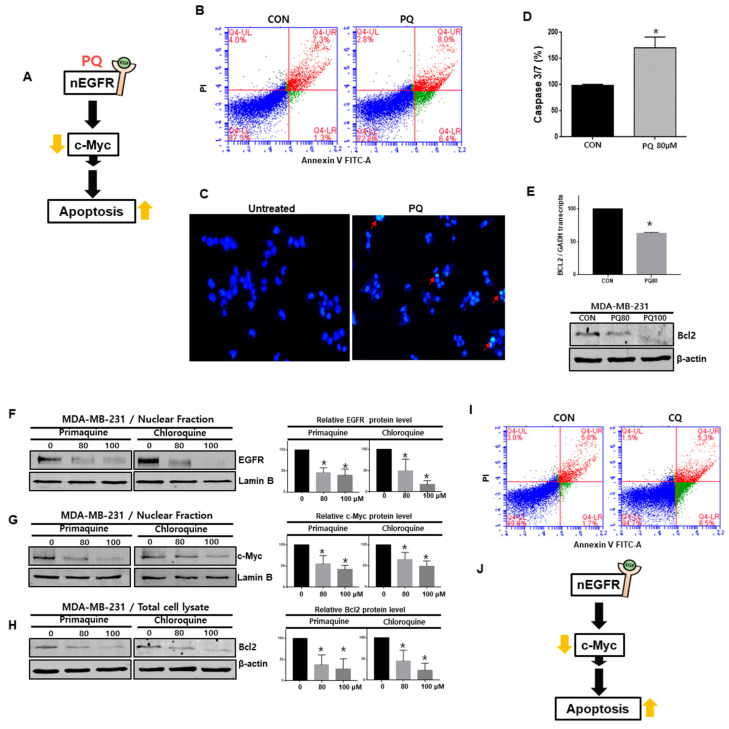
Figure 6.
Primaquine and CQ induced the apoptosis of breast cancer through nEGFR and c-Myc regulation. (A) The proposed model for breast cancer cell death induced by primaquine. (B) Primaquine (80 μM) induced the apoptosis of cancer cells. Cells undergoing primaquine-induced apoptosis were analyzed by using an annexin V-PI staining kit. (C) Analysis of apoptotic cancer cells by fluorescence staining. The nuclei of breast cancer cells were stained with Hoechst 33258 (magnification, ×100). The red arrows indicate apoptotic bodies. (D) The caspase-3/7 activity of breast cancer cells was determined with the Caspase-Gloss 3/7 kit. (E) Effect of primaquine (0, 80, and 100 μM) on Bcl-2 protein levels in breast cancer cells. (F–H) Effect of primaquine and CQ (0, 80, and 100 µM) on nEGFR, cMyc and Bcl-2 protein levels in breast cancer cells. (I) CQ (80 μM) induced the apoptosis of cancer cells. Cells undergoing CQ-induced apoptosis were analyzed by using an annexin V-PI staining kit. (J) The proposed model for CQ-induced breast cancer cell death. The data are presented as the mean ± SD; n = 3; * indicates p < 0.05 vs. control.

