Figure 1.
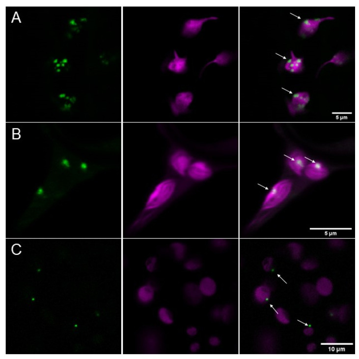
AtPII aggregates in focal structures in chloroplasts. (A) AtPII-GFP (green) under the control of p35S (p35S CaMV::AtPIIcDNA-GFP) and co-expressed with mCherry-tagged transit peptide of tobacco Rubisco (CD3-999 pt-rk [24]; magenta) localizes to plastids in transiently transformed N. benthamiana 2 days after infiltration. (B) Genomic AtPII-GFP (green) expressed under the control of the endogenous PII promoter (pAtPII::AtPIIgenomic-GFP) and co-expressed with mCherry-tagged transit peptide of tobacco Rubisco (CD3-999 pt-rk; magenta) localizes to plastids in transiently transformed N. benthamiana 2 days after infiltration. (C) Genomic AtPII-GFP (green) under the control of endogenous pAtPII (pAtPII::AtPIIgenomic-GFP) localizes to plastids (magenta) in stably transformed A. thaliana. In each row, the GFP fluorescence is shown first, the mCherry fluorescence is shown second, and the merge of both pictures is shown last. White arrows mark exemplarily AtPII aggregates.

