Figure 6.
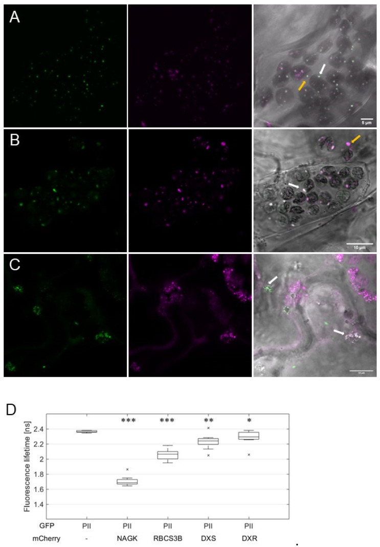
AtPII is found in different plastidial aggregates. AtPII-GFP was co-expressed with AtRBCS3B-mCherry (A), AtDXR-mCherry (B), and AtDXS-mCherry (C), respectively, under the control of p35S in N. benthamiana. In each row, the GFP fluorescence is shown first, the mCherry fluorescence is shown second, and the merge of both fluorescence images with the brightfield image as background is shown last. White arrows mark exemplarily AtPII aggregates in chloroplasts (dark and round structures in the brightfield image), orange arrows indicate extraplastidic vesicle-like structures. (D) FLIM analyses of fluorescent co-localizing signals in (A–C) together with AtPII-GFP/AtNAGK-mCherry as positive control. Student’s t-test used for calculation of significance. Data points marked with an “x” represent statistical outliers of measurement. * p < 0.05; ** p < 0.01; *** p < 0.001.

