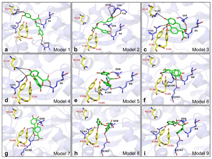
Figure 7.
Docking results of RA binding to the Fe3+ binding site of VmFbpA. (a–i) Docking models 1–9 after running docking simulation using AutoDock (version Vina 1.1.2). RA is shown as sticks in green. Fe3+ binding site of VmFbpA (N138, Y195, and Y196) shown as sticks in yellow. The amino acid residues predicted to form hydrogen bonds toward RA are shown as sticks in purple. The hydrogen bonds predicted by PDBePISA are shown as black dashed lines. VmFbpA is shown as a cartoon with a transparency of 60% and colored in purple.

