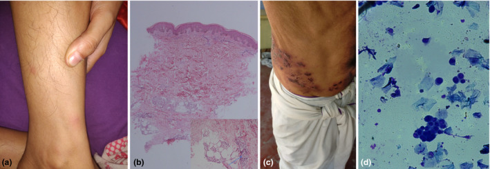
Figure 1.

(a) Multiple erythematous nodules over the leg. (b) Histopathology of lesion revealing lymphoplasmacytic infiltrate around deep dermal vessels and infiltration of subcutaneous fat by lymphomononuclear cells and a few neutrophils (arrow). (c) Closely grouped vesicles coalescing to form bullae and erosions with haemorrhagic crusting in the right T10 dermatome in Case 2. (d) Tzanck smear demonstrating presence of acantholytic cells and multinucleate giant cells (May‐Grünwald Giemsa stain; original magnification: 1000×).
