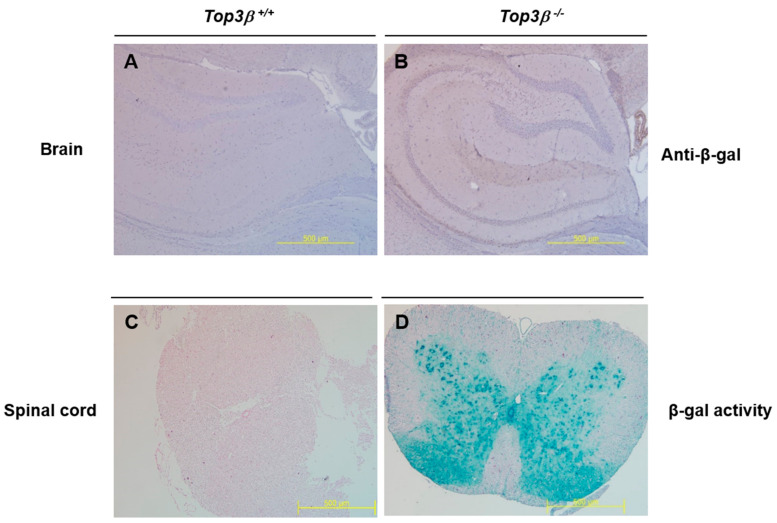
Figure 6.
Top3β gene expression is clear in the central nervous system. Brain tissue block was cut into 4 μm thicknesses and subjected to immunohistochemistry staining. Meyer’s hematoxylin was used as a counter stain. (A,B) Immunohistochemistry staining with antibody against β-galactosidase shows abroad positive signal in the brain of Top3β−/− mouse (×40). (C,D) X-gal staining in the full spinal cord of Top3β−/− mouse shows clear β-galactosidase activity in Top3β−/− mouse (×40).

