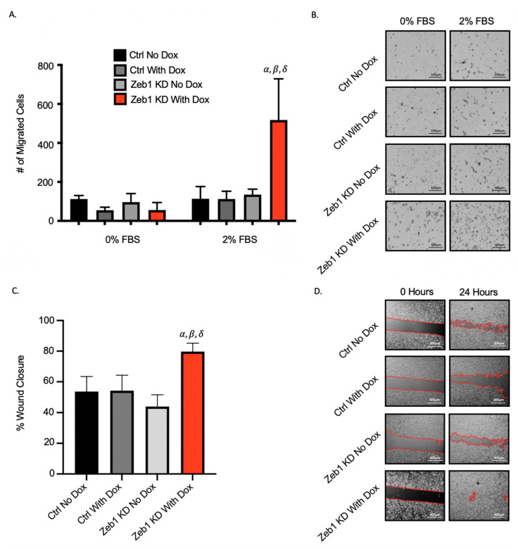
Figure 2.
Knockdown of Zeb1 in PC-3 prostate cancer cells increases cell migration. (A,B) Transwells were coated with 6 µg/well of gelatin. Cells (5 × 10⁴/well) were added to wells and either control media (0% fetal bovine serum [FBS]) or chemoattractant media (2% FBS) was added and cells were allowed to migrate for 18 h. Cells were fixed with 1% glutaraldehyde and mounted with DAPI-containing mounting media. (C,D) For physical barrier wound healing assays, cells were seeded and grown to 90–100% confluency. The physical barrier was removed and cells were allowed to migrate into the wound for 36 h. Representative images are shown for each assay; with migration calculated based on 5 high-powered fields of view (HP-FOV) per well. Black scale bars = 100µm, white scale bars = 300µm. Data is presented as the mean ± standard error of the mean (SEM) (n = 3). α = significantly different than control (ctrl) no doxycycline (Dox). β = significantly different than ctrl with Dox. δ = significantly different than Zeb1KD (Zeb1 knockdown) no Dox (p ≤ 0.05).

