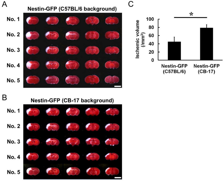
Figure 2.
TTC staining of Nestin-GFP mice (C57BL/6 background) (A) and Nestin-GFP mice (CB-17 background) (B) tissue at 1 day after MCAO. The ischemic areas of Nestin-GFP mice (C57BL/6 background) were rarely observed in the posterior regions of the cortex; they occasionally ranged from the cortex to the striatum (No. 2 and No. 4 in A). In contrast, Nestin-GFP mice (CB-17 background) exhibited reproducible ischemic areas, which ranged from the anterior to the posterior cortex regions (B). Based on these results, the infarct volume of Nestin-GFP mice (C57BL/6 background) was significantly smaller than that of Nestin-GFP mice (CB-17 background), and the coefficient of variation was greater in Nestin-GFP mice (C57BL/6 background) than that in Nestin-GFP mice (CB-17 background) (C). Scale bars: 5 mm (A,B). * p < 0.05 between the two groups of mice [Nestin-GFP mice (C57BL/6 background) vs. Nestin-GFP mice (CB-17 background)] (C) (n = 5 for each group). Abbreviations: GFP, green fluorescent protein; TTC, 2,3,5-triphenylteterazolium; MCAO, middle cerebral artery occlusion.

