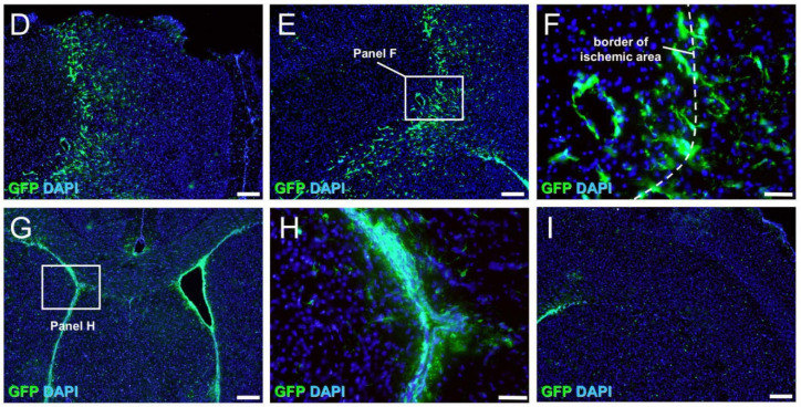
Figure 4.
Immunohistochemistry for GFP at 3 days after MCAO (A–I). GFP was distributed at the site of the ischemic areas, including the peri-ischemic areas (B–F), as well as in the SVZ (G,H), whereas GFP was rarely observed in the contralateral cortex (I). [GFP (B–I: green), DAPI (B–I: blue)]. Results are representative of three replicates. Scale bars: 200 µm (B,D,E,G,I) and 50 µm (C,F,H). Abbreviations: DAPI, 4′,6-diamidino-2-phenylindole; GFP, green fluorescent protein; MCAO, middle cerebral artery occlusion; SVZ, subventricular zone.


