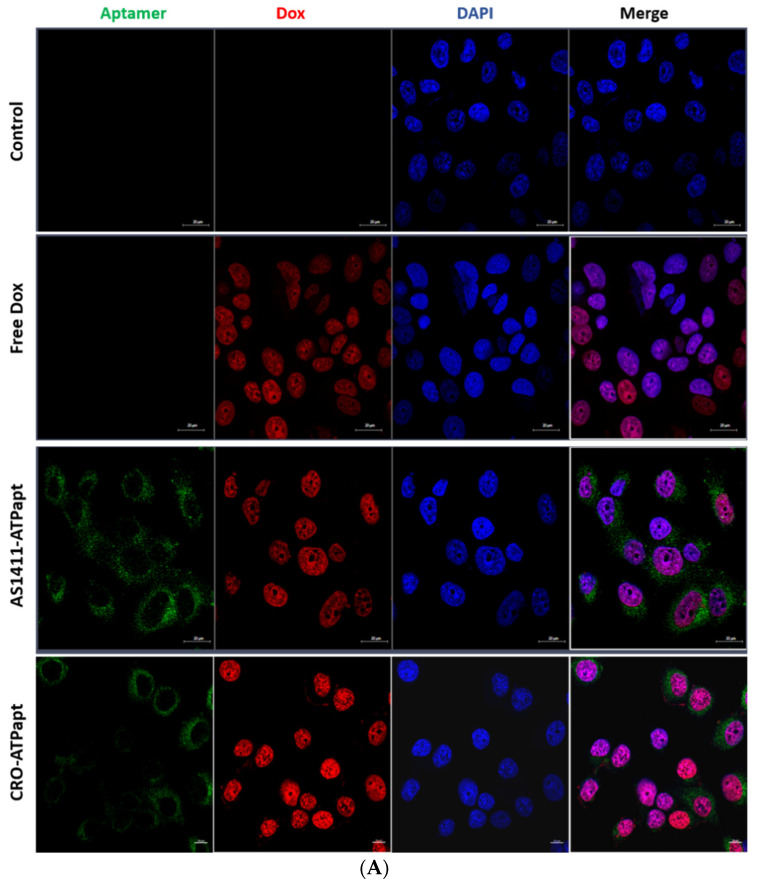
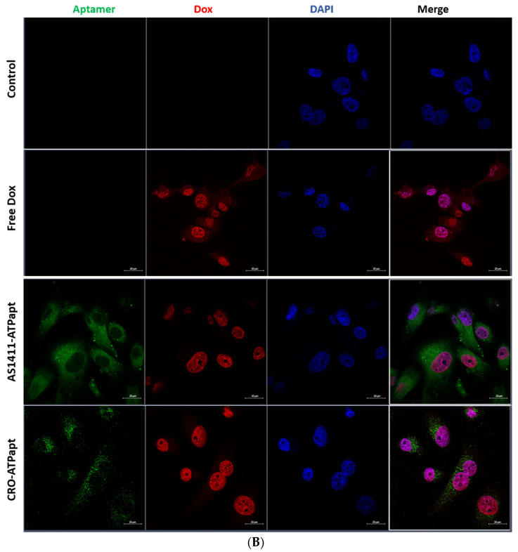
Figure 8.
Cellular uptake of chimeras demonstrated by confocal microscopy. The cancer cells (A) MCF-7 and (B) U87 were incubated with a culture medium containing free doxorubicin or doxorubicin loaded into fluorophore-labeled chimeras. For negative control, cells were incubated with complete culture medium without treatment. After incubation at 37 °C for 3 h, the cells were fixed in 4% formaldehyde and then analyzed by confocal laser microscope (Dox showed red fluorescence, Cy5-labeled ATP aptamer showed green fluorescence, and the nuclei stained with DAPI showed blue fluorescence).


