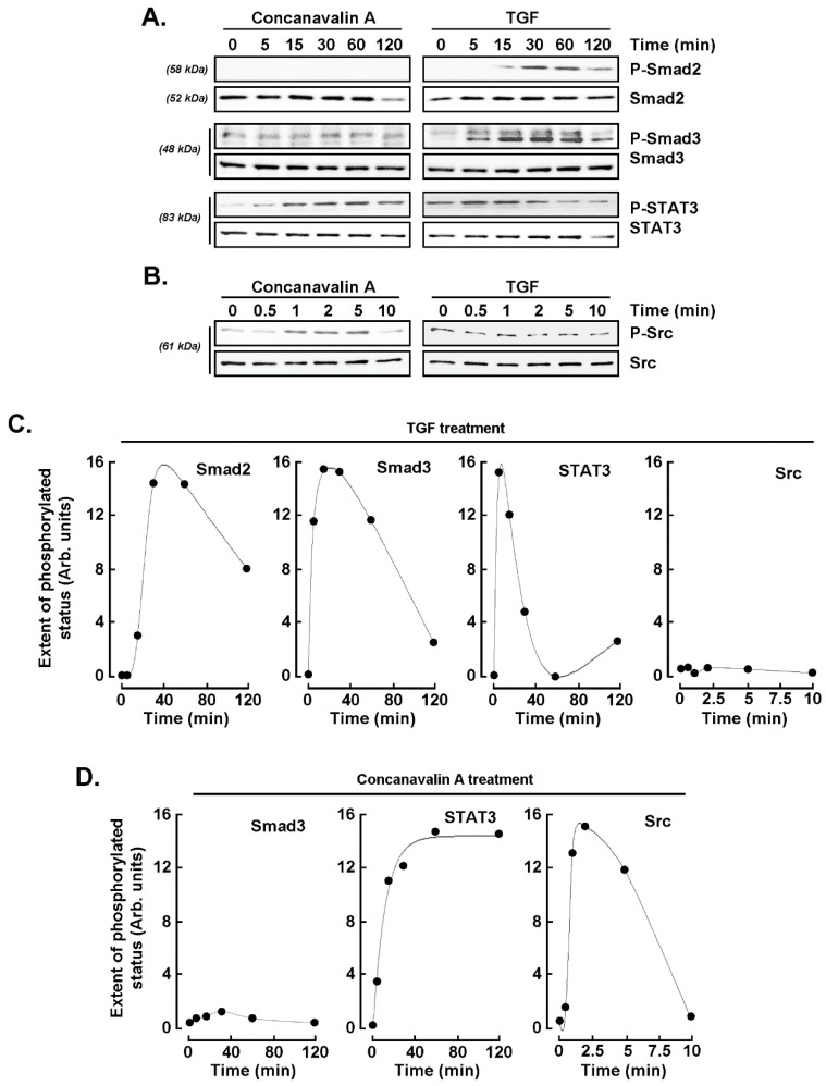
Figure 5.
Concanavalin A and TGF-β share common signaling axis. U87 glioblastoma cells were treated with 10 nM TGF-β or 30 μM ConA for the indicated times, and cell protein lysates were harvested. Long time-course was performed from 0 to 120 min in (A) to monitor Smad2, Smad3, and STAT3 protein status, whereas a short time-course was performed from 0 to 10 min in (B) to monitor Src phosphorylation status. Scanning densitometric analysis was performed of a representative experiment for (C) TGF-β treatment and (D) concanavalin A treatment.

