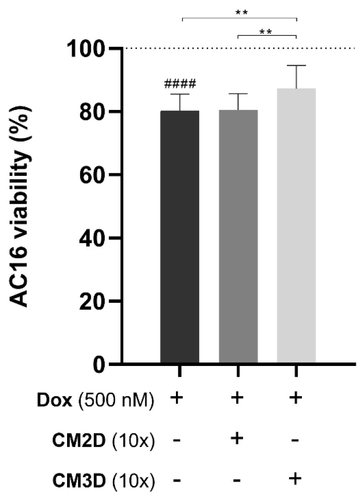
Figure 6.
Exposure of AC16 cardiomyocyte cells to CM3D increased the viability of cells exposed to Dox. The differentiated AC16 cells were incubated with 500 nM of Dox alone or in combination with CM2D or CM3D being the cytotoxicity evaluated by MTS reduction assay. Cell viability is expressed in percentage (mean ± SD, n = 3–6) to non-treated differentiated AC16 cells (control). Grid line represents 100% cell viability (control). Statistical significance is expressed relative to Dox as ** p < 0.01 and to control of non-treated cells, i.e., 100% viability as #### p < 0.0001. CM2D, conditioned medium derived from 2D cultures; CM3D, conditioned medium derived from 3D cultures; Dox, doxorubicin.

