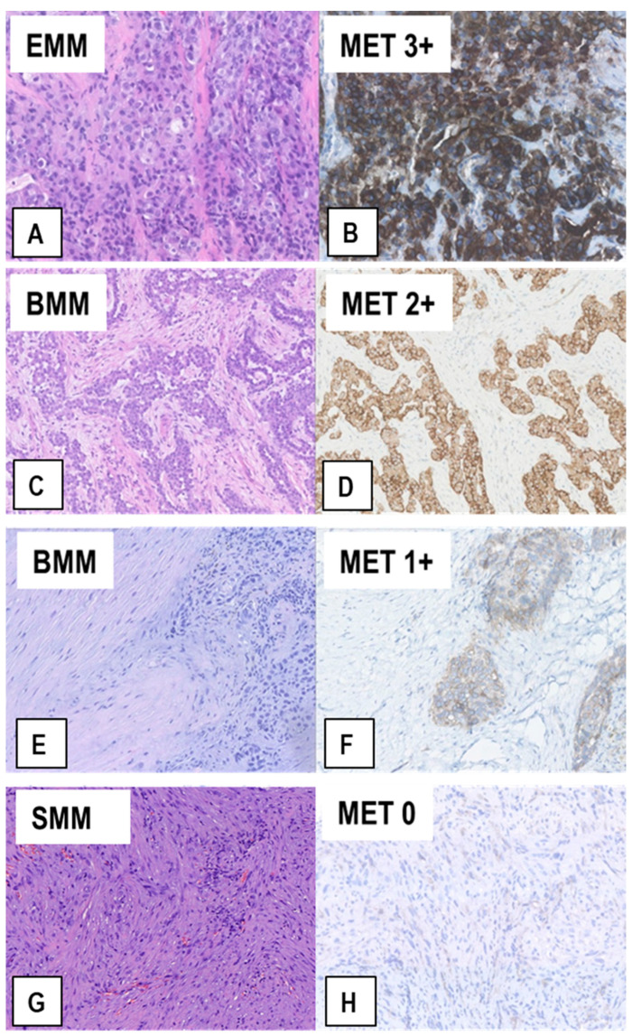
Figure 3.
Representative examples of MET expression in tissue sections from MM of different histological types. Serial sections from each case were stained with H&E (A,C,E,G) and immunostained with anti-MET SP44 mAb (B,D,F,H). In (A,B), an EMM showing strong upregulation of MET expression (MET immunoscore 3+). In (C,D), a BMM with moderately upregulated MET expression (MET immunoscore 2+), which is only present in the epithelioid component. In (E,F), a BMM with weak MET expression (MET immunoscore 1+), which is only present in the epithelioid component, whereas the sarcomatoid component is negative. In (G,H), a SMM with no MET expression (MET immunoscore 0), in which scattered reactive endothelial cells with quite weak MET expression can be observed. (Magnification: (A,B), ×320; (C–H) ×200).

