Figure 1.
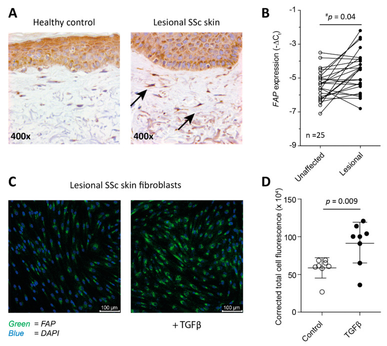
(Myo)Fibroblasts in lesional SSc skin express FAP. Skin biopsies stained for FAP using IHC show presence of FAP positive fibroblasts (black arrows) in lesional SSc skin (A). On average, FAP gene expression is significantly increased in lesional SSc skin compared to unaffected skin of the same patients as determined by a one-sided paired Students’ T-test. The reported # p value is a corrected p value, the uncorrected p value is 0.0013 (B). Primary fibroblasts obtained from lesional SSc skin express FAP in vitro as detected by immuno-fluorescence microscopy (C), and FAP expression is significantly increased by stimulation with 10 ng/mL TGFβ for 5 days (D).

