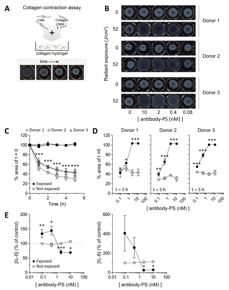
Figure 4.
Application of FAP-tPDT in 3D culture of primary lesional SSc skin fibroblasts cultured in collagen type 1 hydrogels. Primary skin fibroblasts of 3 different donors were seeded in collagen type 1 hydrogels (A) and exposed to a dose response of tPDT after 48 h. Subsequently contraction was visualized after 5 h (B) and quantified over time for a dose of 10 nM PS-antibody construct. In hydrogels not exposed to light, contraction occurs but this is fully blocked by tPDT (C). Dose response curves for each individual donor after 5 h are depicted in (D) (all n = 4 replicates per donor). IL6 and IL8 levels were measured in the supernatant of the hydrogels 5 h after tPDT procedure (E). (Pooled data from duplicates of all 3 donors). * = p < 0.05, ** = p < 0.01, *** = p < 0.001.

