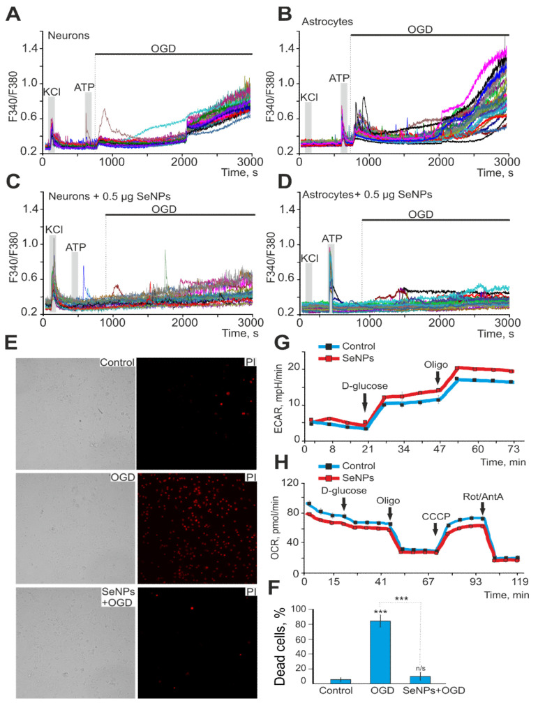
Figure 5.
Protective effect of 0.5 μg/mL SeNPs from OGD-induced [Ca2+]i increase and necrosis. (A,B) Ca2+-signals of neurons (A) and astrocytes (B) during 40 min OGD. (C,D) Ca2+-signals of neurons (C) and astrocytes (D) during 40 min OGD after 24 h incubation with 0.5 μg/mL SeNPs. (E) Images of cortical cell culture in transmitted light and propidium iodide fluorescence detection channel in control (without OGD), after 40 min OGD (OGD) and 24 h treatment with 0.5 μg/mL SeNPs (SeNPs + OGD). The white dots represent the PI-stained nuclei of necrotic cells. (F) Average number of PI-stained cells that died in control due to OGD-induced necrosis in the absence of SeNPs (OGD) and after 24 h incubation with SeNPs (SeNPs+OGD) (% ± SE). Short-term applications of 35 mM of KCl and 10 µm of ATP were used to detect neurons and astrocytes, respectively. Statistical significance was assessed using paired t-test. n/s—data not significant (p > 0.05), *** p < 0.001. (G,H) Effect of 2.5 μg of SeNPs on the rate of acidification of the extracellular medium ECAR (G) and the rate of oxygen consumption OCR (H) in astrocytes after the application of 10 mM D-glucose, 4.5 μM of oligomycin, and a mixture of 2.5 μM of rotenone and 4 μM of antimycin A.

