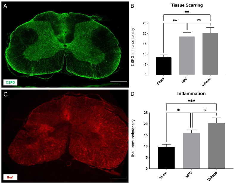
Figure 5.
Tissue scarring and inflammation in the injured spinal cord eight weeks after severe cervical SCI. (A) Spinal cord cross-section of an NPC animal stained for CSPG, a marker for proteoglycans in the extracellular matrix at 10× magnification (scale bar = 500 µm). (B) The immuno-intensity of the CSPG-staining and thus the extent of tissue scarring showed no significant difference between NPC animals (group 1) and Vehicle animals (group 2; n = 8 animals per group; one-way ANOVA with Tukey-HSD-test; p = 0.8281). (C) Spinal cord cross-section of a Vehicle animal stained for Iba1, a marker for macrophages at 10× magnification (scale bar = 500 µm). (D) The immuno-intensity of the Iba1-staining and thus the number of Iba1+ macrophages was not significantly reduced in NPC animals compared to Vehicle animals (n = 8 animals per group; one-way ANOVA with Tukey-HSD-test; p = 0.1468) (* p < 0.05; ** p < 0.01; *** p < 0.001 and ns = not significant).

